The app provides a training tool for medical professionals and students to learn bronchial anatomy. Users can navigate airway segments, identify lymph node stations, and confirm location with real CT images.
by Sharp Vision Software, LLC★ 3.02 ratingsMedicalFreeData updated 2026-10-20
Calculate using TNM classification system. Input tumor size, lymph node involvement, and distant metastasis. Provide summary based on input. Offer educational information about staging criteria.
Provides a color-coded reference for identifying stations on CT scans. It includes anatomic definitions, N-category information, and sampling options. Users can access mediastinal, low cervical, and hilar stations with clear localization guidance.
View 3D models of the , including benign tumors, cancer, lymph node metastasis, and tracheal invasion. Explore relationships between the and surrounding structures like the trachea, nerves, and blood vessels.
Tool allows users to input tumor size, lymph node involvement, and metastasis data to determine stage groupings for non–small cell and small cell cancers using the 9th Edition TNM Classification. Results are displayed based on clinical guidelines.
View detailed anatomical images with CT, MR, X-ray, ultrasound, and 3D modules. Navigate through sectional views and compare with cadaveric structures.
Access references, anatomical diagrams, and clinical calculators. View imaging examples and use decision support tools. Calculate dosages and interpret findings. Share notes and images with colleagues.
The app provides anatomy drawings and allows users to create custom references by adding and editing descriptions for labels on images. It also includes flashcards for Human Anatomy topics and supports iPad natively.
The app provides a detailed guide to musculoskeletal structure, function, and common injuries. It features thousands of 3D models for visualization and learning about anatomy, movement, and pathologies.
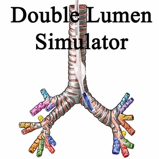
The app offers a simulator for double lumen endotracheal tube placement. It provides real bronchoscopy video images and allows users to practice DLT and bronchial blocker positioning without time constraints.