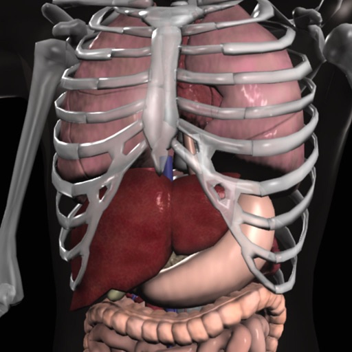
Explore the human body with detailed 3D models of all systems including skeletal, muscular, nervous, cardiovascular, respiratory, digestive, reproductive, lymphatic, and connective tissues.
Patients record audio of urinations within the . Machine learning measures rate and volume. Results are available in a secure provider portal for physicians.
The app provides an interactive view of the head and brain using augmented and virtual reality. Users can explore layers from skin to brain structures and animate brain areas.
Review and explore images of on radiographs, organized by category. Search for quickly. Identify common and uncommon equipment. Access detailed information on device appearances.
Learn histology with interactive slides and quizzes. Explore 19 main topics and 148 subtopics. Identify 2179 slides based on photomicrographic images. Test knowledge with built-in assessments. Access content offline without internet connection.
Create and manipulate geometric shapes such as points, lines, angles, and conic sections. Adjust properties and calculate metrics. Display shapes on a zoomable coordinate system. Construct medians, altitudes, and bisectors.
Access detailed information including eight common types, treatment approaches, and documentation guidelines. View clinical images and case studies. Receive updates and references for evidence-based practices.
Access medical protocols for specific EMS agencies. View detailed guidelines for emergency procedures. Retrieve password-protected agency protocols from Training Officers. Includes references and supporting materials for each protocol set.
Capture and document visible light images and video of patient conditions. Select clinical information from menu options. Submit images to Centricity Clinical Archive for storage and viewing.
Monitor energy levels by measuring specific acupuncture points. Identify organ and tissue conditions based on electrical resistance. Track progress over time with detailed reports. Share results with healthcare professionals for further evaluation.
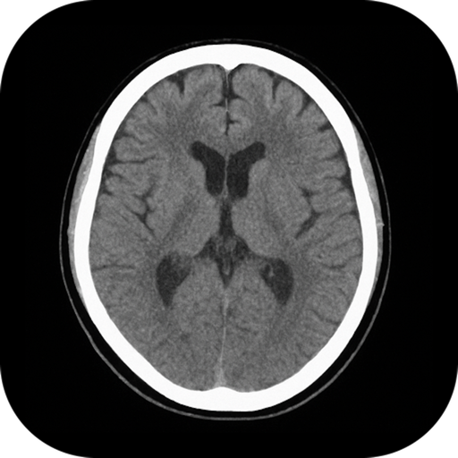
View and analyze medical images with support. Load and browse files quickly. Use 3D preview, MPR, and image fusion for detailed analysis. Measure, annotate, and compare images. Store and share findings locally without internet access.
The app provides a learning experience about organs using 3D models, video, audio lectures, quizzes, and text. It covers detailed information on various organs, cells, tissues, and organ systems with diagrams and interactive elements.
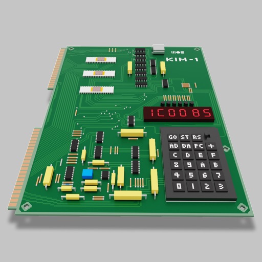
Users can assemble and debug assembly code using a simple interface with 7-segment LEDs and buttons. Run programs on an emulated 8-bit processor. Solve programming puzzles and explore hardware components used in early home computers.