Use a virtual microscope to examine samples and identify tissue types. Cut samples from succulent, onion, celery, and elodea plants. Recognize xylem, phloem, sclerenchyma, collenchyma, parenchyma, apical , stomata, and cuticle.
Explore 3D models of DNA, cells, and plant tissues. Perform virtual dissections to reveal labels and definitions. View interactive simulations and animations for biological processes.
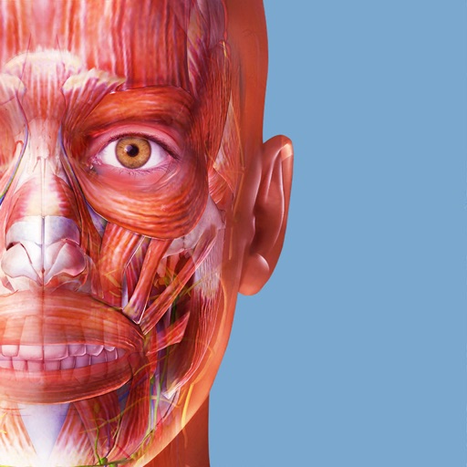
Explore human anatomy and physiology through interactive 3D models. Rotate, zoom, and dissect virtual organs. Access detailed explanations of systems and diseases. View layered structures and compare healthy vs. pathological states.
The app provides a detailed guide to musculoskeletal structure, function, and common injuries. It features thousands of 3D models for visualization and learning about anatomy, movement, and pathologies.
Explore with interactive 3D models of male and female bodies. View cadaver and diagnostic images alongside 3D structures. Access key organ views and animations. Purchase additional physiology and dental content.
The app presents microscope slide images of vertebrate histology and mitosis in fish embryo. Text describes tissue functions and locations, covering the four basic tissue types.
The app presents microscope slide images of vertebrate histology and mitosis in fish embryo. It describes tissue functions and locations, serving for review and learning with interactive practice sections.
Learn anatomical structures through and quizzes. Study with beginner-friendly content. Track progress and set learning goals. Review key terms and definitions. Test knowledge with interactive challenges.
Explore 3D human anatomy models with interactive touch controls. Rotate and zoom for detailed views. Perform virtual dissections to reveal muscle layers and anatomical structures. Take 3D location quizzes to test knowledge.
Test your knowledge with trivia questions about human and medicine. Learn about body systems, organs, and biological functions. Track progress with results and review correct answers. Share scores with friends.
Explore 3D anatomical models with interactive rotation and labeling. View detailed cross-sections and layers. Access structured learning modules and quizzes. Save custom study notes and share annotations.
Review histological micrographs to test knowledge and identify structures. Access high-quality images from the University of Michigan Virtual Slide collection. Use 27 sets covering over 550 pages of tissues, organs, and organ systems.