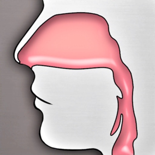
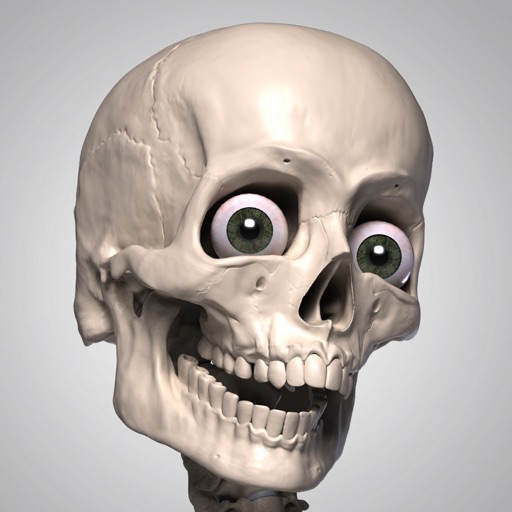
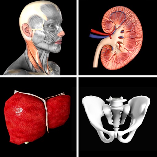
The app provides a 3D simulator for exploring the upper airway. Users can view detailed information on 34 anatomical structures and navigate the space with various controls.
Explore poses with science-based anatomy explanations. View 3D interactive models of body structures. Learn biomechanics of each asana. Track progress and set practice goals. Access expert insights from Dr. Gill Solberg.
Study canine acupoints with detailed illustrations.
This app assists canine acupuncturists in studying, reviewing, and practicing acupoints. It covers 361 transpositional and 77 classical acupoints with detailed descriptions, indications, and pictures.
Explore 3D models of ligaments and skeletal systems. Rotate, zoom, and dissect virtual structures. Access detailed labels and anatomical notes. Upgrade to unlock additional systems and features. View cross-sectional views and compare structures.
The app assists canine acupuncturists in studying, reviewing, and practicing acupoints. It covers 361 transpositional and 77 classical acupoints with detailed descriptions, indications, and multiple illustrations.
Patients can mark pain areas on a 3D model to identify affected muscles. Therapists can view muscle anatomy, referred pain patterns, and associated symptoms. The includes a free version with 12 muscles and an option to purchase the full version.
The app provides animated anatomical illustrations and videos to teach normal swallowing structure. It includes lateral, posterior, and superior views with structure identification by touch, zoom, and pan capabilities.
Learn through multiple-choice quizzes on 416 structures or image matching. Create custom quizzes or focus on missed topics. Access 3D renderings of bones, muscles, and organs. Set a timer for timed challenges. Track progress with results.
Create and study using an enhanced spaced repetition algorithm. Review cards based on performance and difficulty. Collaborate on with others in real time. Access the across multiple devices through a web version.
The app provides practice organizing intra-oral radiographs for dental professionals. Learners can arrange 18 to 20 individual images in the correct orientation for diagnostic purposes.