
Plant Histology Lite
Carolina Biological Supply
Microscope images of plant cell division.
The app presents microscope slide images of plant mitosis and meiosis. It includes interactive practice sections for learning and review.

Set illumination on your microscope
Learn illumination setup through interactive simulation. Adjust microscope controls and observe image changes in real-time. View control locations and step-by-step explanations. Practice setting up illumination for accurate microscopy.
Showing 1–20 of 1,000 ranked alternatives.
Carolina Biological Supply
Microscope images of plant cell division.
The app presents microscope slide images of plant mitosis and meiosis. It includes interactive practice sections for learning and review.

Motic China Group Co., Ltd
Connect, view, capture, and measure.
The app connects to Moticam X cameras and Moticam or Digital Microscopes. Users can view, capture, annotate, share, and measure images from their microscope.



Thermo Fisher Scientific
View images, share, and explore protocols
Access fluorescence microscopy resources including image galleries, protocol details, and material lists. View full-screen images with descriptions and spectral data. Share images easily and provide feedback through an in- button.

John Sader
Calibrate cantilevers
Calibrate atomic force microscope cantilevers using the . Input resonant frequency and quality factor measurements. Calculate spring constant and display results. Export data for analysis.

elklab UG (haftungsbeschraenkt)
Explore the microscopic world with magnification.
The app transforms a mobile device into a microscope for viewing small objects with magnification. Users can capture images of revealed details and share discoveries.

Carolina Biological Supply
Microscope slide images of animal histology.
The app presents microscope slide images of vertebrate histology and mitosis in fish embryo. It describes tissue functions and locations, serving for review and learning with interactive practice sections.

Carolina Biological Supply
Microscope slide images of animal tissues.
The app presents microscope slide images of vertebrate histology and mitosis in fish embryo. Text describes tissue functions and locations, covering the four basic tissue types.

Carolina Biological Supply
Plant cell division reference and learning.
The app presents microscope slide images of plant mitosis and meiosis. It allows comparison of monocots and dicots, with macroscopic views included.


Jiovany Orozco
Identify with microscope
Use a virtual microscope to examine samples and identify tissue types. Cut samples from succulent, onion, celery, and elodea plants. Recognize xylem, phloem, sclerenchyma, collenchyma, parenchyma, apical , stomata, and cuticle.


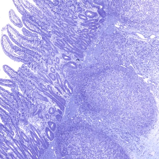
Eolas Technologies Inc.
Explore slides in detail
View ultra-high-resolution images of tissue samples. Zoom in and pan across microscopic slides. Access detailed annotations and labels. Search for specific tissues or structures. Compare images side-by-side.
Apps are ranked by functional similarity, category fit, rating confidence, and popularity.