Assessing cardiac structure and function visually.
This medical application shows normal blood flow within the heart using two and three-dimensional echocardiography. It demonstrates standard views with rotatable heart models and video clips for assessing cardiac structure and function.
by University Health Network★ 3.02 ratingsMedical$4.99Data updated 2026-10-23
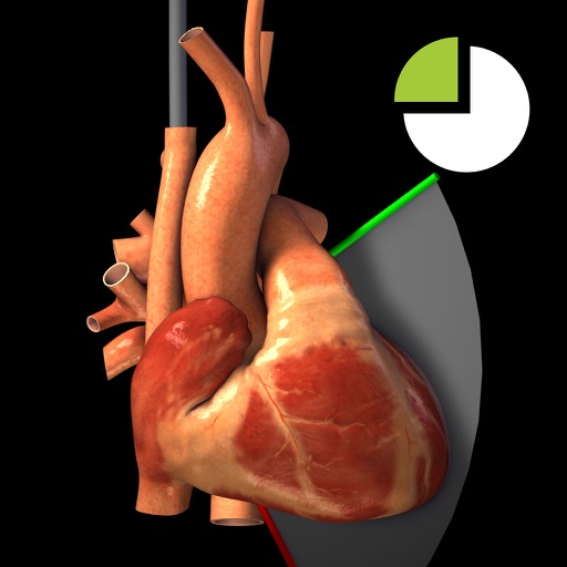
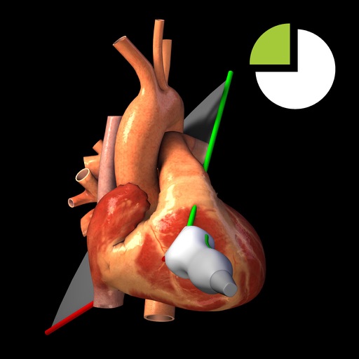
View TEE recordings for 20 diagnostic and corresponding 3D heart models. See TEE probe position and ultrasound plane alignment. Assess cardiac function through interactive visualizations.
Users can view TTE recordings for 20 cardiac and interact with corresponding 3D heart models. Rotate and adjust 3D models to visualize cardiac structures. Annotate and compare images with reference guidelines.
The app provides a learning environment to explore 19 non-standard TEE views. It uses a rotatable 3D heart model and associated TEE clips to help visualize complex 3D heart structures.
Medical reference for echocardiography specialists.
The app is a medical reference focused on echocardiography. It includes a searchable index of specialized topics and a database of figures, images, and videos related to echocardiography findings.
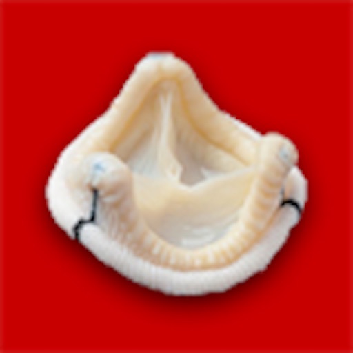
The app supports echocardiography professionals in assessing prosthetic cardiac valve function. It offers guidelines for evaluating implanted valves and selecting valves before implantation based on published research.
The app allows users to record EKGs with Kardia devices, share heart data with doctors, and track health history. It provides immediate analysis of normal, possible atrial fibrillation, bradycardia, or tachycardia.
The app supports neonatologists in training programs for neonatal echocardiography. It provides videos and loops to familiarize users with basic echocardiography views for self-directed learning.
The app provides an interactive learning tool for Focused Cardiac Ultrasound (FOCUS) assessments. It displays transthoracic echocardiography recordings alongside rotatable 3D models of the heart and probe for five standard views.
Process and quantify perfusion studies of the heart, kidney, liver, and skeletal muscle using contrast-enhanced ultrasound. Align images automatically or manually. Color-code digitally-subtracted images.
Learn Transesophageal Echocardiography with interactive lessons and a question bank. Access a cognitive aid for emergency scenarios. Log and submit cases to the NBE. View offline loops and pathology examples.
Explore the structure and function of the through interactive 3D models. Learn about IgAN and C3 glomerulopathy with detailed explanations. Track symptoms and progress with personalized notes.
Draw and paint images using customizable color pencils, pastels, and brushes. Choose colors freely and adjust settings for precise control. Save artwork to camera roll and share via Facebook or Twitter.
Provides visual guides for 28 . Users can navigate through detailed images, learn associated structures, and access descriptions. The includes interactive features for quick reference during procedures.
Access real patient heart and lung sounds for auscultation practice. Listen to and compare heart and lung sounds. Complete modules with quizzes and feedback. Track progress and review explanations for each sound.
Provides information on heart defects in neonates and infants. Includes descriptions of cardiac defects, diagnostic criteria, and imaging findings. Offers guidance for learners in echocardiography.
This app presents animated content on performing echocardiographic studies. It covers cardiac anatomy, transducer positions, and image acquisition for professional lectures.