The app provides a training tool for medical professionals and students to learn bronchial anatomy. Users can navigate airway segments, identify lymph node stations, and confirm location with real CT images.
by Sharp Vision Software, LLC★ 3.02 ratingsMedicalFreeData updated 2026-10-20
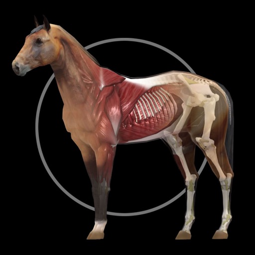
The app presents a 3D model of horse anatomy, including muscles, bones, and organs. It features detailed models of the hoof, head, and pelvis, with labeled structures and audio pronunciations.
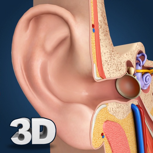
Explore a 3D anatomical model with 360° rotation, zoom, and camera movement. Slice anatomical parts and detailed structures. Take notes and access labeled part names in English offline.
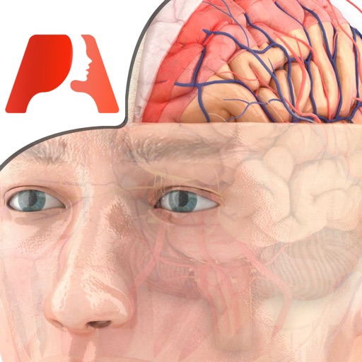
Explore 3D neuro-anatomy models with layered views and cross-sectional animations. View nerve pathway diagrams and study detailed anatomical structures. Access over 50,000 words of educational content on neurological topics.
Study with 1500+ exam-like questions covering anatomy, kinesiology, pathology, and techniques. Use flashcards to reinforce memory. Access exams to prepare for certification tests.
Access cardiology content and unlock full features
Medical "Access cardiology patient care guidelines, physical exam techniques, and EKG interpretation. View symptom assessments and basic to advanced clinical content. Unlock full features with in- purchase."
Access high-resolution anatomical images with pinch-zoom and label toggling. View interactive digital version of Netter's Atlas of Human . Purchase in- to unlock full content. Includes drug guides, calculators, and ICD 10-CM resources.
Patients can mark pain areas on a 3D model to identify affected muscles. Therapists can view muscle anatomy, referred pain patterns, and associated symptoms. The includes a free version with 12 muscles and an option to purchase the full version.
View 3D anatomy models and interactive animations of endovascular devices by pointing the camera at a flat surface. Explore device technology through visual demonstrations. Share content for educational purposes.
The app presents detailed medical illustrations, CT scans, and 3D rendered images of wrist anatomy. It includes a series of cases demonstrating hand and wrist pathology.
The app presents human anatomical systems and their physiology through 3D animation. Each system is detailed in a chapter with life-like animations and narration for understanding function.
Learn about and valve anatomy through detailed illustrations. Explore pathophysiology of valvular disease. Review surgical techniques for valve repair and replacement. Access educational content for physicians and medical students.