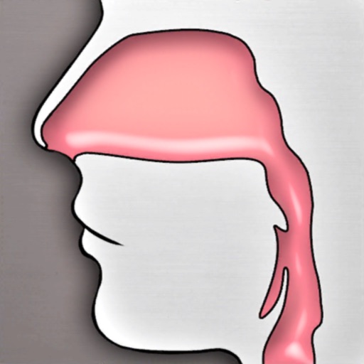
The app provides a 3D simulator for exploring the upper airway. Users can view detailed information on 34 anatomical structures and navigate the space with various controls.
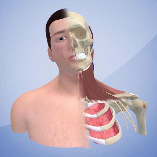
Explore 3D models of anatomy. Rotate postures 360 degrees for detailed views. Adjust head rotation and tilt. View comprehensive muscle layers. Simulate head tremors. Scan an OR code to activate the model from a workbook.
Explore 3D models of ligaments and skeletal systems. Rotate, zoom, and dissect virtual structures. Access detailed labels and anatomical notes. Upgrade to unlock additional systems and features. View cross-sectional views and compare structures.
Study with interactive meridian pathways and point locations. Explore a customizable anatomy model with muscles, bones, and organs. Edit and save detailed descriptions of points, meridians, and models. Search by point name, function, or indication.
Access detailed human anatomy images for study and reference. Explore over 26,000 medical images with interactive 3D views. Search for specific anatomical structures. Annotate and bookmark images for later review.
Explore detailed 3D models of skeletal, blood, muscular, nervous systems, and male and female organs. Use virtual dissection to identify organ positions. Access over 500 articles for in-depth study.
Explore a realistic 3D model of the human with adjustable lighting and background. Manipulate the model for full range of motion using dynamic sliders. Switch between user-defined poses and view layered anatomical structures.
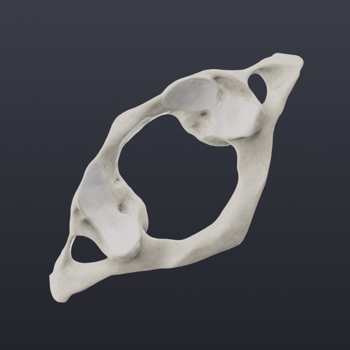
Explore 3D models to study nerves, vertebrae, and cranial structures. Rotate and zoom to examine anatomical details from multiple angles. Watch animations explaining muscle contractions.
Retrieve anatomical structures and clinical data instantly
Explore anatomical structures and clinical details with instant retrieval. Search over 1000 anatomical items including ligaments, tendons, arteries, and organs. Browse by system layers such as skin, skeletal, and circulatory.
View detailed anatomical images with CT, MR, X-ray, ultrasound, and 3D modules. Navigate through sectional views and compare with cadaveric structures.
Review and analyze radiographs and CBCT scans to identify anatomical structures, dental disease, and technical errors. Test knowledge with quizzes on radiographic anatomy and image quality.