Access product information, view surgical techniques, and review training materials. Download product brochures and specifications. Schedule and manage sales appointments. Track inventory and order status. Share content with colleagues and clients.
The app presents detailed medical illustrations, CT scans, and 3D rendered images of wrist anatomy. It includes a series of cases demonstrating hand and wrist pathology.
Assess wounds with automated measurement and image analysis. Capture and store patient data securely. Share images with healthcare providers for remote consultation. Track healing progress over time. Generate reports for clinical documentation.
Ask medical questions to get structured responses. Review clinical guidelines and literature summaries. Attach images or PDFs for context. Organize clinical reasoning with structured outputs. Streamline documentation and decision-making processes.
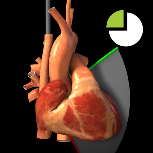
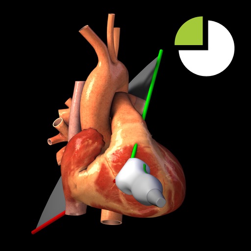
View TEE recordings for 20 diagnostic and corresponding 3D heart models. See TEE probe position and ultrasound plane alignment. Assess cardiac function through interactive visualizations.
The app provides access to medical images and reports on the Nuance PowerShare Network. Users can upload clinical images from their iOS device for secure sharing with medical professionals.
Manage health conditions with personalized care plans, set medication reminders, and communicate with healthcare providers. Track symptoms, view treatment progress, and access educational resources for managing chronic conditions.
Users can view TTE recordings for 20 cardiac and interact with corresponding 3D heart models. Rotate and adjust 3D models to visualize cardiac structures. Annotate and compare images with reference guidelines.
Process patient data to support diagnostic decisions. Analyze medical history, lab results, and imaging. Provide synthetic knowledge and dynamic reference data. Offer analytics for treatment planning and patient care guidance.
Manage IVF lab operations with AI-powered quality control tools. Upload and analyze embryo images for competency assessment. Track lab workflows, set alerts, and access open-source image databases for training and evaluation.
This atlas educates radiologists on normal and pathological bony anatomy of the foot and ankle. It utilizes interactive 3D models and illustrations for a comprehensive understanding of musculoskeletal CT interpretation.