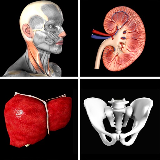
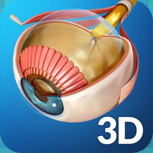
Explore through interactive 3D models. Tap labeled structures for detailed explanations. Switch between anatomical systems with intuitive controls. Review lab notes and learn at your own pace.
The app presents all 268 pages of Leonardo da Vinci's anatomical drawings from the Royal Collection. It features a beautiful interactive book interface with pinch-zoomable, high-resolution images and a magic spyglass to decode mirror writing.
Explore interactive 3D models of reptiles, amphibians, tide pools, human anatomy, and the solar system using augmented reality. Tap to interact with models, view animations with sound, and access detailed content from printed materials.
Explore human through interactive 3D models. Point device at AR or demo marker to view skeletal, respiratory, digestive, circulatory, and urinary systems. Identify body parts and learn about their functions.
Diagnose and manage orthopaedic trauma with interactive decision flows. View hundreds of diagrams and illustrations. Access evidence-based, clinically relevant information for trauma and orthopaedics.
Learn through multiple-choice quizzes on 416 structures or image matching. Create custom quizzes or focus on missed topics. Access 3D renderings of bones, muscles, and organs. Set a timer for timed challenges. Track progress with results.
The app presents detailed medical illustrations, CT scans, and 3D rendered images of wrist anatomy. It includes a series of cases demonstrating hand and wrist pathology.
Explore an interactive 3D model to view muscles, bones, and joints in detail. Rotate, zoom, and dissect layers to study human movement. Access expert insights on biomechanics and . Track progress with customizable study plans and quizzes.
Explore points and meridian pathways with interactive models. Adjust anatomical structures by hiding or fading. Edit and save detailed descriptions of points, meridians, and anatomy. Search points by name, function, or indication.
View cross-sectional CT scans of head, neck, chest, and abdomen. Navigate through slices, adjust window levels, and measure distances. Annotate images and save or share findings. Supports anatomic localization and future modality expansions.
Patients can mark pain areas on a 3D model to identify affected muscles. Therapists can view muscle anatomy, referred pain patterns, and associated symptoms. The includes a free version with 12 muscles and an option to purchase the full version.