This atlas educates radiologists on normal and pathological bony anatomy of the foot and ankle. It utilizes interactive 3D models and illustrations for a comprehensive understanding of musculoskeletal CT interpretation.
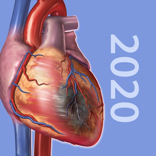
View 3D models of human and pathologies. Rotate, zoom, and examine structures from any angle. Explore detailed digestive system models and rare diseases. Access medically accurate information for learning and reference.
Learn and review point of care with labeled videos, illustrations, and still images. View anatomy diagrams and exam examples. Access pathology content for diagnostic reference. Perform virtual exams and track progress through interactive modules.
Access a library of images that speak in a voice for common words and phrases. Tap icons to hear words used in daily interactions like greeting, calling, dining, and healthcare communication.
The app offers a collection of pictures and videos for users to speak with a female voice. It includes common words and phrases for various situations and videos demonstrating tongue and lip movements for key words.
Learn anatomy through x-ray, CT, MRI, and ultrasound images. Identify normal structures and common pathologies. Practice clinical reasoning and test imaging test selection. Track progress with structured curriculum based on training.
The app presents videos of vocal pathologies including small, medium, large, and hemorrhagic polyps. It includes audio clips of patients reading the rainbow passage to demonstrate the effect of each disorder on vocal fold function.
Practice board-style questions with detailed rationales and high-yield explanations. Track progress with practice quizzes and -style simulations. Review comprehensive content to prepare for the 1 .
Access detailed diagrams and clear explanations for science topics. Watch animated, narrated video tutorials with adjustable playback speed. Take notes, highlight key points, and save progress. Sync across devices for continued learning.