Explore 3D anatomical models with interactive navigation. View over 8200 structures. Rotate, zoom, and dissect virtual organs. Access detailed labels and cross-sectional views. Compare male and female . Save custom views and annotations.
by 3D4Medical.com★ 0.00 ratingsMedical$22.99Data updated 2026-09-08
Explore 3D models of male and female anatomy with interactive views of all body systems. Search for anatomical structures and layers including skeletal, muscular, and circulatory systems. View detailed information on each system and its components.
This atlas educates radiologists on normal and pathological bony anatomy of the foot and ankle. It utilizes interactive 3D models and illustrations for a comprehensive understanding of musculoskeletal CT interpretation.
Create and customize , take , and practice with tests. utomated study schedules. Access community-made and purchase professional study packs for specific subjects. Track progress and review weak areas with AI-driven insights.
Learn anatomy through x-ray, CT, MRI, and ultrasound images. Identify normal structures and common pathologies. Practice clinical reasoning and test imaging test selection. Track progress with structured curriculum based on training.
Explore through interactive 3D models. Tap labeled structures for detailed explanations. Switch between anatomical systems with intuitive controls. Review lab notes and learn at your own pace.
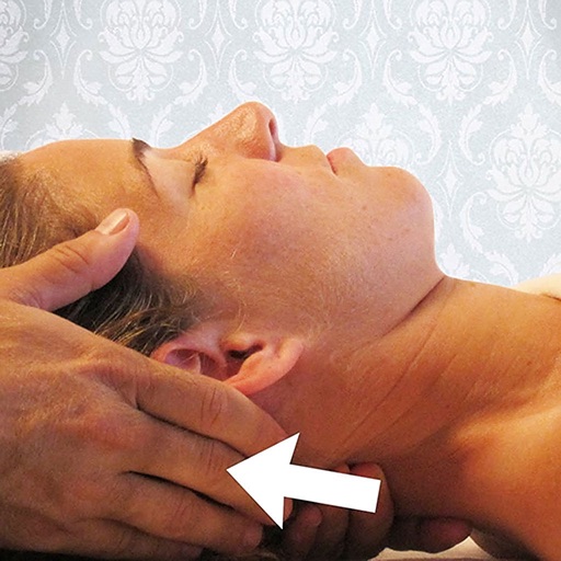
Learn with video demonstrations, still images, and text descriptions. Browse by body area or alphabetically. Access 13 full routines ranging from 10 to 60 minutes. Practice for specific conditions like back pain or athlete recovery.
Learn and occlusion with interactive 3D models and detailed explanations. Study tooth morphology, identify structures, and practice quizzes. Save progress and review notes for exam preparation.
Diagnose and manage orthopaedic trauma with interactive decision flows. View hundreds of diagrams and illustrations. Access evidence-based, clinically relevant information for trauma and orthopaedics.
View 3D anatomy models and navigate through layers. Draw annotations, animate physiological processes, and illustrate medical conditions. Share annotated images and export interactive content for educational use.
Patients can mark pain areas on a 3D model to identify affected muscles. Therapists can view muscle anatomy, referred pain patterns, and associated symptoms. The includes a free version with 12 muscles and an option to purchase the full version.
Explore detailed 3D anatomical models of the human body. Rotate, zoom, and dissect virtual organs. Access interactive tutorials and quizzes. Annotate and save notes. Share models with colleagues or students. Export 3D views for presentations.