The app simulates a ripple tank, allowing users to create waves with their finger. It demonstrates wave phenomena like interference, diffraction, refraction, resonance, and the Doppler effect.
Access -ray data for elements. Search for elements by symbol or name. View electron configuration and levels. Export data as PDF or text. Share information via email or messaging.
The app provides details on preparation, positioning, and an exposure guide for taking diagnostic X-rays of horses. Each view includes an example X-ray from a normal horse.
Capture patient images with guided steps. Adjust settings for optimal clarity. Generate detailed reports for patients. Streamline workflow to save time and reduce equipment costs. Improve treatment explanations to boost business.
Explore 3D anatomical models with interactive layers and systems. View structures by body parts or organs. Access over 6,000 labeled anatomical objects. Study for exams and tests. Navigate through realistic human body visualization.
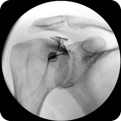
The app enables physicians to access radiological images on a mobile device. It offers secure communication features, including messaging and VoIP, for sharing images and information.
Medical imaging device Capture and analyze ultrasound images in real time. Use AI for instant diagnostic support. Store, share, and export scans. Integrate with workflow software for seamless data management.
Explore human with 3D models of muscles, bones, blood vessels, nerves, and organs. Rotate, zoom, and dissect layers to view internal structures. Access detailed explanations and MRI-based data for each system.
Enables medical professionals to prioritize acute cases using AI-driven alerts. Notify teams of critical findings and share imaging results instantly. Access real-time notifications and collaborate on urgent decisions within the .